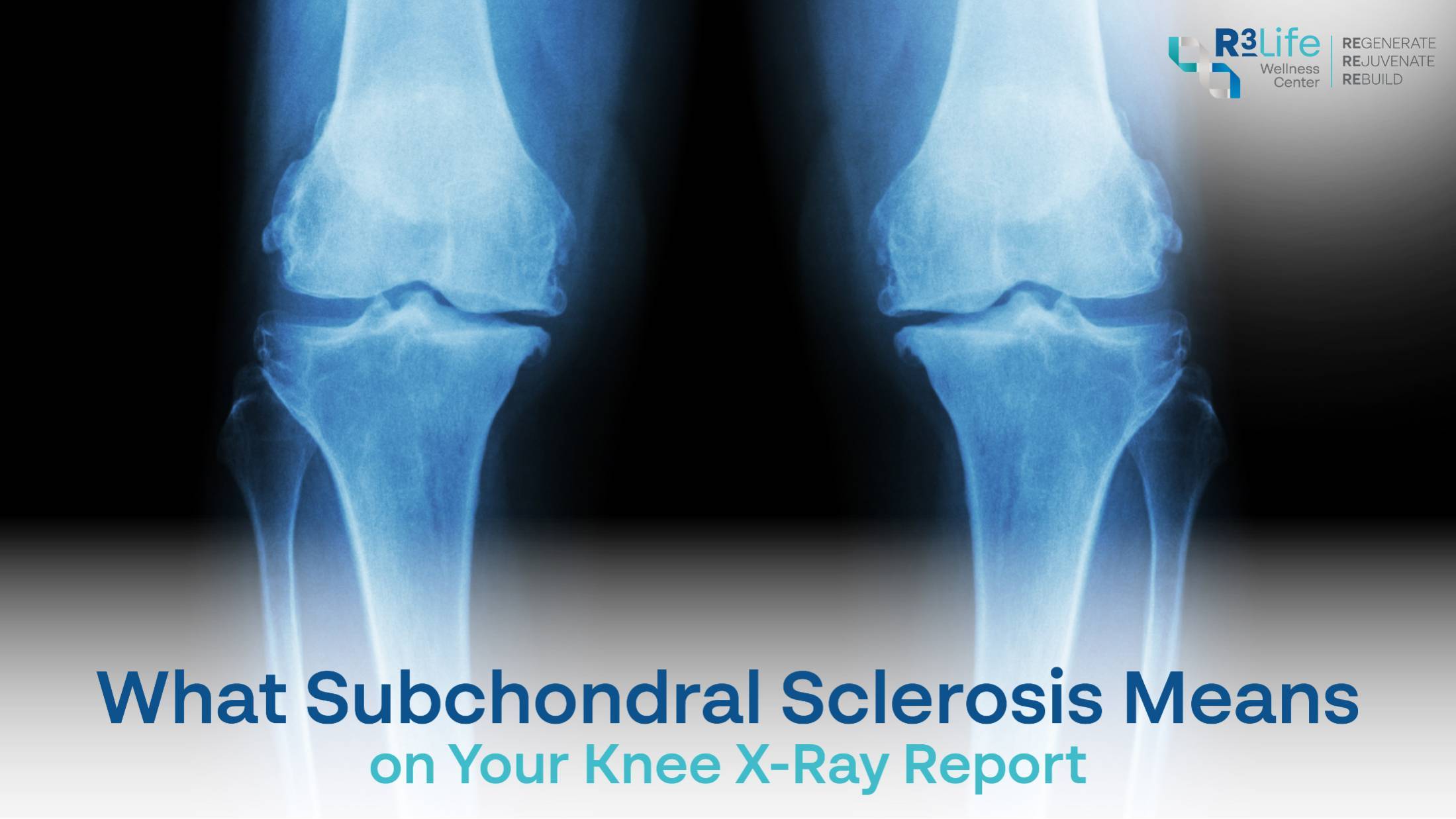
What Subchondral Sclerosis Means on Your Knee X-Ray Report
Key Highlights
- Subchondral sclerosis describes thickened, denser bone directly beneath the cartilage, and it appears as a brighter white band on an X-ray.
- It is a radiographic finding rather than a diagnosis, which means it describes what the bone looks like and nothing more.
- The bone thickens because it is absorbing more load than it used to, usually as the cartilage above it thins.
- Imaging severity and symptom severity correspond poorly, so a report that reads badly does not reliably predict how a knee will feel.
- Exercise-based conservative care remains the first-line approach in every major osteoarthritis guideline, regardless of what the scan shows.
Quick Summary
You went in about a knee that has been aching for months. You came out with a report containing a phrase nobody explained, and a search history that now includes the word "sclerosis."
Subchondral sclerosis sounds far more ominous than it is. It is one of the standard things a radiologist notes when reading a joint X-ray, it is extremely common, and on its own it says nothing about how much pain you should expect or what treatment you need.
This article explains what the term actually describes, why it appears, why imaging and symptoms so often fail to agree, and what usually happens next.
What Subchondral Sclerosis Is
Breaking the term into its parts makes it considerably less alarming than it appears on a report.
The Bone Beneath the Cartilage
"Subchondral" simply means beneath the cartilage. It refers to a thin plate of bone sitting directly under the smooth cartilage surface that lines the joint, along with the honeycomb-like bone underneath that.
"Sclerosis" means hardening or increased density. Put together, subchondral sclerosis describes bone in that specific location that has become thicker and denser than it previously was. Nothing in the term implies infection, tumor, or damage in the way patients often assume when they first read it.
Why It Shows Up White on an X-Ray
X-rays work by measuring how much radiation different tissues absorb. Denser material absorbs more and appears whiter, which is why bone shows up brightly and soft tissue barely registers.
When the subchondral bone becomes denser, it absorbs more radiation than the surrounding bone, and a radiologist sees a brighter, more sharply defined band along the joint surface. The whiteness on the film is the density itself, appearing exactly as any increase in bone density would.
Sclerosis Is a Finding, Not a Diagnosis
This distinction is the single most useful thing to take from the report. A finding describes an observation; a diagnosis explains what is causing a problem and guides treatment.
Subchondral sclerosis is one of several features radiologists look for when assessing a joint, alongside narrowing of the space between bones, bone spurs, and small fluid-filled cavities. Those features together may support a diagnosis of osteoarthritis, but any one of them in isolation is a descriptive observation waiting for clinical context.
Why It Appears
Sclerosis is not something that happens to bone at random. It is a response, and understanding what it is responding to explains most of what patients want to know.
How Load Transfers When Cartilage Thins
Healthy cartilage distributes force across the joint surface and cushions the bone beneath it. As cartilage thins, more of that force reaches the subchondral bone directly, and it reaches concentrated areas rather than being spread evenly.
Bone is living tissue that remodels according to the demands placed on it. Loaded more heavily, it responds by laying down additional minerals and becoming denser. Subchondral sclerosis is therefore an adaptive response, essentially the bone reinforcing itself where it is working hardest.
The Link With Osteoarthritis Staging
Because it tracks with cartilage loss, sclerosis is one of the features used in the standard radiographic grading systems for osteoarthritis. It typically appears alongside joint space narrowing, which serves as an indirect measure of remaining cartilage.
It is worth knowing that these grading systems describe structure only. A grade is a description of what the joint looks like on film, and it was never designed to predict how much pain someone experiences or how they will respond to treatment.
Other Causes Worth Ruling Out
Osteoarthritis is much the most common explanation, but it is not the only one. Increased subchondral density can also follow:
- A previous fracture or significant joint injury
- Disrupted blood supply to a region of bone
- Inflammatory forms of arthritis, which have a different underlying mechanism
- Repetitive high-impact loading, particularly in athletes
This is one reason the report is read alongside your history and examination rather than on its own. The same appearance on film can arise from different processes, and they are managed differently.
What It Does and Does Not Tell You
Knowing the limits of the imaging is what stops a report from causing more distress than the knee itself.
What Imaging Can Show
A plain X-ray is genuinely useful for a defined set of questions:
- The space between the bones, as an indirect indicator of cartilage thickness
- The presence and size of bone spurs
- The density and contour of the subchondral bone
- Overall alignment of the joint and how load is distributed across it
What it cannot show is equally worth knowing. Cartilage itself is invisible on X-ray, as are the ligaments, the joint lining, and inflammation within the joint. It also cannot show pain, which has no radiographic appearance whatsoever.
Why Imaging and Symptoms Often Disagree
This is the part that unsettles patients most, and it is thoroughly documented in the research. A substantial proportion of people with clear radiographic osteoarthritis report no knee pain at all, while others with mild-looking films are significantly limited by their symptoms.
The likely explanation is that structural change on film and the experience of pain are driven by overlapping but different things. Inflammation of the joint lining, changes within the bone marrow that only appear on MRI, fluid in the joint, and how the nervous system processes persistent pain signals all contribute, and none of them is visible on a standard X-ray.
The practical consequence is that your report should be interpreted next to your symptoms, never instead of them.

What Usually Happens Next
For most people the finding does not change the treatment plan. It confirms a picture rather than opening a new question.
Questions Worth Asking Your Doctor
A short list makes the follow-up appointment considerably more productive:
- Which compartment of the knee is affected, and is the joint space narrowed there
- Does this finding change what you would recommend, or confirm what you already suspected
- Is my alignment loading one side of the joint more than the other
- Is there anything in the report that points away from osteoarthritis
- Would further imaging change the plan, or would it just add detail
That last question is worth asking directly. Additional imaging is valuable when it alters a decision and less so when it does not.
Where Conservative Care Fits
Every major osteoarthritis guideline places the same things first, and they are not the things patients expect. Structured exercise and strengthening carry the strongest evidence of anything available, alongside weight management where relevant, because reducing load through the joint addresses the mechanism directly.
Where symptoms persist despite that foundation, R3 Life Wellness Center offers established conservative options at the Silom–Surawong flagship branch. Ostenil Plus is a hyaluronic acid viscosupplement injected into the joint to lubricate it and absorb shock. PRP (Knee) uses a preparation made from your own blood, and may support the healing of tendons, ligaments, and surrounding tissue while easing osteoarthritis pain.
Neither replaces the exercise foundation. They sit alongside it.
When Regenerative Options Are Discussed
Mesenchymal stem cell therapy tends to enter the conversation when conservative measures have been given a fair trial and symptoms remain limiting, but surgery is either unwanted or not yet appropriate.
The honest position is that current evidence points most consistently toward improvements in pain and function rather than toward rebuilding lost cartilage, and claims about cartilage regrowth run well ahead of what has been demonstrated. R3 Life Wellness Center's Cord Tissue-MSCs and Amnion-MSCs are consultation-led therapies, and the appropriate starting point is an assessment of whether the option makes sense for your particular joint and stage. Outcomes differ among individuals.
Frequently Asked Questions about Subchondral Sclerosis
Q: Is subchondral sclerosis serious?
A: On its own, no. It is a common radiographic finding describing denser bone beneath the cartilage, and it is most often an adaptive response to increased load. Where it does carry weight is in grading: when osteoarthritis is present, subchondral sclerosis is one of the features radiologists use to classify how advanced the changes are, alongside joint space narrowing and osteophytes. Even then, what matters clinically is your symptoms and function, not the appearance of the bone on film.
Q: Does subchondral sclerosis mean I have osteoarthritis?
A: Not by itself. It is one of several features that together may support an osteoarthritis diagnosis, alongside joint space narrowing and bone spurs, and no single finding confirms or excludes the condition on its own. Its absence is equally uninformative, as early-stage osteoarthritis often shows other changes before any sclerosis appears. Subchondral sclerosis can also follow injury, inflammatory arthritis, or disrupted blood supply, which is why it is read alongside your history and examination.
Q: Can subchondral sclerosis be reversed?
A: The bone density change is not something current treatment sets out to reverse, and reversing it is not the goal. Treatment targets the load and the symptoms, and people frequently improve substantially in pain and function while the radiographic appearance stays unchanged.
Q: My X-ray looks bad but my knee feels fine. Should I be worried?
A: This is common and well documented. Many people with clear radiographic osteoarthritis have no pain at all. It is worth staying active and maintaining strength around the joint, but a film that reads worse than your knee feels is not a reason to restrict what you are doing.
Q: Do I need an MRI as well?
A: Often not. An MRI shows cartilage, ligaments, and bone marrow that an X-ray cannot, but it is most useful when the result would change the plan, such as when a mechanical problem like a meniscal tear is suspected. Ask your doctor directly whether it would alter their recommendation.
Conclusion
Subchondral sclerosis is one of the more alarming-sounding phrases a patient can find on an imaging report and one of the less alarming things it can describe. The bone beneath the cartilage has thickened in response to carrying more load than it used to, which is an adaptation rather than damage. It is a finding, not a diagnosis, and it needs your symptoms and your examination beside it before it means much of anything.
Assessment at R3 Life Wellness Center is led by physicians holding ABAARM board certification in anti-aging and regenerative medicine, with certification in cell therapy from the Academy of Cell Therapy Thailand. To discuss an imaging report or a joint that is not improving with conservative care, visit r3lifewellness.com or message +66 88 689 8888 on WhatsApp.

 Benefits of Stem Cells: How does it help to restore the body, and can it really be used to treat diseases?